


If an external force with changing amplitude acts on an elastic medium such as a gas, a liquid or a solid, an undulating propagation of pressure and density fluctuation occurs in space and time, starting from the point where the force is applied. This is known as sound. The frequency of sound waves ranges from a few hertz (Hz) up to several gigahertz (Figure 1).
© PI | https://www.pi-usa.us/en/
Infrasound, the sound humans cannot hear, lies at frequencies below 16Hz. It’s followed by the hearing range, which reaches up to 20kHz. Ultrasonic waves, which cannot be heard, lie in the frequency range from 20kHz to 1.6GHz, which equals 16 billion cycles per second. A prominent application example in medical technology is the use of ultrasound for diagnostic imaging techniques. In industry and research, ultrasound is mainly used in measurement technology, where sound waves with low power are used. The intensity of the sound describes the power that hits a certain surface. If it exceeds 10W/cm2, we speak of power ultrasound, which often shows frequencies in the range of 20kHz to 800kHz.
Power ultrasound in medicine
Piezo ceramics generate power ultrasound by using the piezoelectric effect to transform high frequency electric energy into mechanical and vice versa. The ultrasonic waves generated by a piezoelectric transducer can be used in fluids to generate so-called cavitation bubbles. These are created when acoustic sound waves generate rarefaction and compression zones with locally higher and lower material densities in a medium. The resulting high tensile and shear forces between the zones cause the fluid medium to be ripped apart creating gas-filled cavities.
Depending on the frequency and sound force of the applied ultrasonic sound waves, two different states can occur for the cavitation: 1) a stable cavitation with small oscillating gas bubbles, i.e., they pulsate with the rhythm of the ultrasonic sound frequency and with that increase or decrease in size; and 2) an instable cavitation, where such a high ultrasound power is used that the cavitation bubbles expand so much that they finally implode. In this case, very high forces and pressures are released in the form of shock waves and extremely high temperatures of several 1,000°C, as well as flashes of light can occur temporarily (Figure 2).

© PI | https://www.pi-usa.us/en/
These physical effects of cavitation are used for various medical applications. For example, the stable cavitation is applied in medical imaging when gas-filled microspheres are used as an ultrasound contrast medium. In addition to cavitation, power ultrasound can cause changes in material or even destroy it, which is used, for example, in medical applications, but also in industrial material processing or ultrasonic cleaning.
Therapeutic ultrasound methods
Ultrasound opens new and innovative therapeutic possibilities that improve or even substitute many established procedures: Minimally invasive, and gentle treatment methods with improved therapy success and fewer side effects, can be realized using piezo components.
In this case, therapeutic ultrasound refers to a group of methods in which ultrasound is not only a means of diagnosis but also the core element of therapy itself. By using piezoceramic ultrasonic transducers, minimally- or non-invasive and gentle treatment methods, with improved therapy success, can be realized. These include methods which use focused, but also unfocused, ultrasound – for example tissue ablation, targeted drug delivery, the ultrasound-assisted dissolution of blood clots (thrombolysis), or the fragmentation of kidney stones (lithotripsy). Further application examples using therapeutic ultrasound are cartilage therapy, the administration of drugs through the skin and cosmetic treatments.
Tissue ablation with ultrasound
It is possible to selectively remove certain tissue areas with the help of focused ultrasound – and with a completely contactless procedure (Figure 3). Ultrasound-operated tools make this minimally- or non-invasive surgery technique, which is often also called incisionless, possible today.
The ultrasound-based ablation of tissue, for example the removal of tumors in the prostate or uterus, is carried out extracorporeal and thus non-invasive. For this form of therapy, high-intensity focused ultrasound (HIFU) is projected into the body with the help of piezoelectric elements. The acoustic waves lead to a thermal ablation since the absorption of the ultrasonic wave energy generates heat above 42°C due to friction, so that the proteins of the cell structures denaturize and entire tissue areas within the focus of the sound waves coagulate. HIFU can also be used to treat conditions such as tremor or epilepsy without medication by targeting specific brain structures with heat. Monitoring in these cases is performed simultaneously by magnetic resonance imaging (MRT).

© PI | https://www.pi-usa.us/en/
An alternative method for tissue ablation is histotripsy. In this case, HIFU generates cavitation bubbles, whose high mechanical energy causes cells to burst without any significant heating of the affected tissue.
Targeted drug delivery with sound waves
Ultrasound makes the targeted release or activation of drugs at a specific location in the body possible. Various mechanisms are suitable. The basic method is the injection of microspheres filled with medication or gas, which are distributed in the patient’s bloodstream during treatment and can only be used where they are in the sound field focus. An unstable cavitation generated by ultrasound causes the microspheres to burst in a targeted way. If the small spheres are filled with drugs, active substances can be released locally in the body directly at their destination.
To increase the uptake of therapeutic substances in specific tissues, gas-filled microspheres can be injected. The stable oscillation of these microspheres, which is generated using ultrasound, produces micro streams, which in combination with the mechanical energy of the acoustic waves, increase the permeability, for example, of the blood vessel walls for the active substance in the blood. This mechanism is called sonoporation, and is used, for example in the reversible opening of the blood-brain barrier, a process that can improve the medical therapy of neurodegenerative diseases such as Parkinson’s or Alzheimer’s significantly.
In sonodynamic therapy, focused ultrasound waves induce the formation of reactive oxygen species (ROS) which act cytotoxically on cells, such as, in cancer tumors.
Intravascular therapy with piezo components
An intravascular application of ultrasound is used in a minimally invasive way to reduce atherosclerotic plaques in blood vessels or at the heart valves as well as with life-threatening vascular constrictions (stenosis). In intravascular lithotripsy, ultrasound waves increase the permeability of the blood vessel wall (sonoporation) allowing the medication to more easily penetrate and induce the dissolution of plaques. If a blood clot (thrombus) has already formed, its structure can be loosened with the help of mechanical ultrasound energy. Drugs can then more easily penetrate the thrombus and dissolve it. In this way, the flow of blood to vital organs is ensured and embolisms are prevented.
Why piezoelectric transducers?
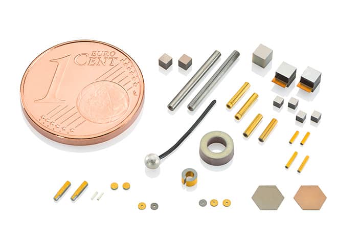
Generating and detecting ultrasound is a classic piezo application of piezoelectric transducers. When an AC voltage is applied, the piezo elements start to oscillate at the frequency used. Ultrasound generation benefits from the short response times and the high dynamics of this motion. Piezo elements (Figure 4) are suitable for various ultrasound applications – especially in power ultrasound since they generate considerable power densities of up to several kW/cm2, which can be applied for kidney stone fragmentation. The advantage here lies in the contactless execution of therapeutic procedures in patients. Ultrasound-operated medical devices are applied extracorporeal, and are, therefore, even less stressful for the patient than established minimally invasive procedures.

© PI | https://www.pi-usa.us/en/
Which transducers are suitable for therapeutic ultrasound?The selection of a suitable transducer for therapeutic ultrasound strongly depends on the specifications of the potential device, such as focus depth, focus size, maximum electrical voltage, installation space, sound pressure and ambient conditions like strong magnetic fields. In the following, various transducer types, which can be used for therapeutic ultrasound, are explained.
HIFU transducer with fixed focus point
Transducers with a device-specific fixed focus point (Figure 5) are mainly used in ultrasonic handpieces, like for cosmetic skin treatments or cartilage therapy. The focus depth, which is usually in the range of a few centimeters, is determined by the shape of the transducer. To generate focused ultrasound, piezoceramic focus bowls can be used. Their radius determines the ultrasound focus depth. Depending on the application, ferroelectric soft or hard ceramics such as PIC255, PIC155, and PIC151 or PIC181 are used.

© PI | https://www.pi-usa.us/en/
The piezoceramic focus bowls can be customized from 2mm to 130mm outer diameter, in various designs and different quantities. Special shape variations are possible – like collar, flattening, phases, spheres, or application-specific designs. The electrode of such a focus bowl can be provided with one or more wraparound contacts to facilitate installation and cable routing in the end device.
HIFU transducer with steerable focus point (phased arrays)
In order to move an ultrasound focus, so called phased array transducers are used. Such ultrasonic transducers are usually made of many piezoelectric elements (array) that can be controlled individually. Using a time-shift electrical control (phase), interferences occur in the sound field, creating a defined and steerable focus point. The motion of the ultrasound focus in these phased array transducers can be controlled by appropriate electronics. Transducers typically used for therapeutic ultrasound are annular phased arrays or curved phased arrays.
Annular phased arrays are group of transducers made of a piezoelectric ceramic disk, ceramic plate, or focus bowl. The applied electrode can be chosen from various material systems, such as Ag thick films, or thin films of Cu or CuNi. Annular phased arrays have a structured electrode that can, for example, be structured using a laser. In this case, the electrode is divided into concentric surfaces in the shape of a central ring with surrounding rings. The width of the electrode rings diminishes from the inner to the outer ring since all electrode segments have the same surface area (Figure 6).

© PI | https://www.pi-usa.us/en
At the front, these annular arrays have an electrode that covers the entire surface. An electronic focus control of annular arrays is possible along the ultrasound beam axis by a phase-shift control of the individual electrodes. Annular arrays are, for example, used in handpieces for prostate tumor ablation.
Curved phased arrays are the most complex sound transducers used for therapeutic ultrasound. They are mainly used for thermal or histotripsy-induced tissue ablation, like of breast cancer, in the abdomen or for the therapy of tremor, as well as epilepsy. To generate a directed ultrasound wave, piezoelectric elements – a few to over several thousand elements depending on the application – are arranged on a concave spherical dome and excited synchronously (Figure 7). This focuses the energy in the shock wave. Commonly used shapes are plates or disks.
Although piezo plates make use of the entire space in the transducer, they still lead to distortions in the sound field and a softening of the sound focus. In contrast, piezo disks have a more homogenous single sound field and are thus better suited for generating the most accurate focusing possible. Special shapes such as pentagons or hexagons are also suitable for lining a HIFU phased array transducer with piezo elements. The electrodes of these sound transducers can e.g. be Ag thick films or thins films of Cu or CuNi.

© PI | https://www.pi-usa.us/en
Structuring the electrode surfaces with wraparound contacts is the best solution to lead any electrical contact to the rear of the transducer. The numerous piezo elements can individually be soldered or glued with stranded wires or cables. Furthermore, customized flexible circuit boards can be used for contacting these complex transducers. The sound focus is controlled in most cases by using the phase offset of the individual signals in the drive electronics used. Concave HIFU phased array transducers are always used in conjunction with a water bellow since this is the only way to overcome the impedance jump between the piezo elements and the surrounding environment with as little loss as possible.
© PI | https://www.pi-usa.us/en
Transducers in minimally invasive catheters
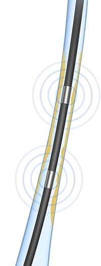
Minimally invasive catheters use ultrasonic sound waves for therapy, like for removal of blood vessel deposits or for targeted tissue ablation for the treatment of atrial fibrillation of the heart (Figure 8).
In this case, individual piezo components, such as miniaturized piezo tubes or plates, are used (Figure 9).
Freely defined shapes with dimensions of only a few millimeters also offer high effective force in a minimal installation space. For transducer applications, it is furthermore possible to assemble several elements in a catheter by soldering or gluing them together. It is important that the piezo elements do not come into direct contact with tissues or bodily fluids. An electrically insulating protective layer made of silicon or parylene, for example, protects against an electrical short circuit and prevents the piezo elements from corrosion.
Therapeutic ultrasound opens numerous new fields of application for the treatment of neurodegenerative, cardiovascular or cancer diseases.
© PI | https://www.pi-usa.us/en


![NBA’s Mental Health Policy Lambasted by Former Player, Royce White [VIDEO]](https://newsfortomorrow.com/wp-content/uploads/2019/08/gettyimages-675633360-225x125.jpg)
