


When it comes to recording and stimulating brain activity, scientists can rely on a formidable tool: light. An international research team, coordinated by IIT- Istituto Italiano di Tecnologia (Italian Institute of Technology), has developed nanometric light modulators that, fabricated on a micrometric optical fiber, make the fiber capable of studying neuronal tissue in deep regions of the brain. The new approach, published in Advanced Optical Materials and featured on the journal’s front cover, lays the groundwork for an innovative type of minimally invasive neural probe that can be used to study the central nervous system. In perspective, the nanomodulators will be employed to study specific brain diseases, including brain tumors and epilepsy.
The study was carried out by IIT in collaboration with the University of Salento (Italy), the Politecnico of Bari (Italy), the Consejo Superior de Investigaciones Cientificas (CSIC, Spain) and the Centro National de Investigaciones Oncologicas (CNIO, Spain).
The study’s first author is Filippo Pisano, a researcher at IIT’s Center for Biomolecular Nanotechnologies (CBN) in Lecce, Italy, coordinated by Marco Grande of the Politecnico di Bari and CBN Principal Investigators Ferruccio Pisanello and Massimo De Vittorio.
In Italy the interdisciplinary team aimed at obtaining micrometric structures which are capable of studying neuronal tissue in a detailed way by using light, i.e. through the incorporation of optical nanomodulators. To do this, scientists combined expertise in nanometer-scale fabrication and biomedical neuro-engineering, in order to exploit the physics of surface plasmon polaritons and create an investigative tool that modifies and amplifies the way light can stimulate and monitor specific brain areas.
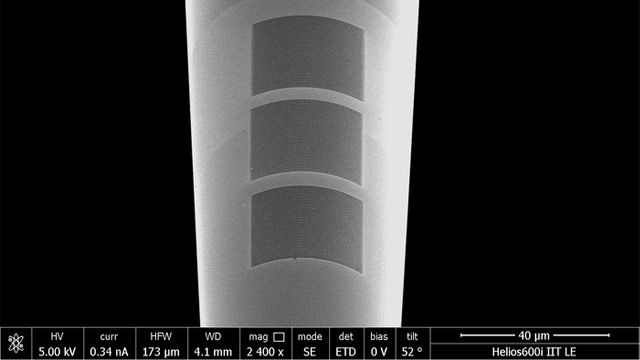
They started from a tapered optical fiber, thinner than a hair, and then they equipped it with nanostructures that resonate in response to a light stimulus introduced by the fiber itself into deep brain regions. The nanostructures were created by coating the probe microscopic tip with a thin layer of gold. Then, using a gallium ion beam as a chisel, they shaped a grid of nanoscopic optical elements, consisting of 100 nm thin lines, whose characteristics were validated in a series of microscopy and optical spectroscopy experiments.
Thanks to this fabrication method, it was possible to obtain a tool which permits to control both the probe light beam modulation and the local electric field acting on surfaces comparable to the size of brain cells. Researchers may be able, then, to study the interaction between the light beam and neuronal structures, even in the deepest areas of the brain.
The possibility of creating such implantable plasmonic systems offers a new perspective in the study of the central nervous system: the amplification produced by the nanostructures is intended to be an efficient tool for detecting the biochemical and cellular structure alterations underlying the origin of several neural disorders.
Therefore, the part of the international team based in Spain is focusing on the application it may have. Experimental researchers at CSIC led by Liset M de la Prida are working to apply these probes in the study of post-traumatic epilepsy and neurodegenerative diseases, such as Alzheimer’s disease. While, the Brain Metastasis Group led by Manuel Valiente at the CNIO will investigate the use of this new technology to distinguish primary from metastatic tumors, whose treatments are different, as well as the use of light to permeabilize the blood-brain barrier, allowing anti-tumour drugs to pass through the vascular barrier.
Reference: Pisano F, Kashif MF, Balena A, et al. Plasmonics on a neural implant: engineering light–matter interactions on the nonplanar surface of tapered optical fibers. Adv. Opt. Mater. 2022;10(2):2101649. doi: 10.1002/adom.202101649
This article has been republished from the following materials. Note: material may have been edited for length and content. For further information, please contact the cited source.


