


WINCHESTER, Va. — Brain and spine surgeries can come down to mere millimeters in terms of being a success or a failure.
At Virginia Brain and Spine Center (VBSC) in Winchester, which partners and works with Valley Health System, new technology is being used so neurosurgeons can perform procedures more efficiently and safely.
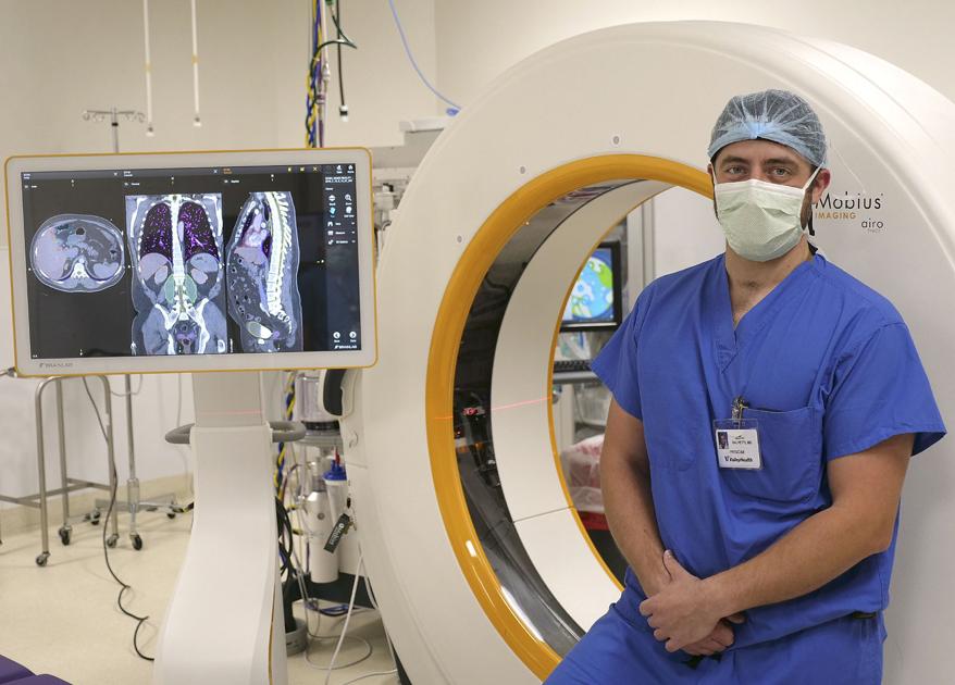
Earlier this fall, Valley Health neurosurgeons began using new technologies that weren’t being used anywhere else in the region. That new technology includes utilizing the Airo Mobile Intraoperative CT and the Curve Image Guided Surgery platform with neuronavigation technology.
The Airo Mobile Intraoperative CT is a CT scanner used in the operating room to provide diagnostic-quality images, giving surgeons up-to-date anatomical information during surgery and allowing for real-time, informed decision-making during the procedure.
The Curve Image Guided Surgery platform displays 3-D images of the patient’s anatomy in an intraoperative position. Curve utilizes a state-of-the-art computer with one or two 26-inch touch-screen monitors and two infrared cameras that track the patient’s exact position on the operating table relative to the surgeon’s instruments.
The two pieces of technology work hand-in-hand as the Airo CT provides real-time feedback to be transmitted and used by the Curve software.
Valley Health acquired the new technology when Dr. David Salvetti joined Valley Health and the VBSC a few months ago. Salvetti said typically when a doctor joins a practice or health system, an investment into new technology is made.
The new technology is used in all of the brain tumor cases as well as all of the cases involving brain functionality but less so for trauma at the center. Spinal surgeons are using it on about 80% of lumbar spinal fusions. It’s not beneficial for all fusions, Salvetti said.
The new systems allow neurosurgeons like Salvetti, who is a general neurosurgeon but has a focus on spinal surgeries, to have a better look at what’s going on around an incision site or an area where something such as a screw is being placed.
“Traditionally, when you’re doing instrumented spinal fusions (or the act of placing a screw into the vertebrae) it’s done with X-Ray. That gives you a one-time snapshot during your surgery of where instruments are positioned and it gives you one plane of view,” Salvetti said.
For example, if a doctor is looking at a patient using a lateral X-Ray, the only thing that can be seen is the lateral X-Ray. There’s no three-dimensional space.
Surgeries become particularly more difficult involving patients with scoliosis, where the spine becomes twisted and distorts a normal anatomy.
Utilizing the scans from the Airo CT and working with the 3-D images and Curve technology, Salvetti can see the surgery from a different point of view while it’s happening.
Typically, the process would begin with exposing the spine as normal , then placing a small array with different reflective markers and scanning the patient with the Airo CT. Then the computer creates a 3-D model with the array as as reference point and a camera that looks at the array and decides which instruments have similar arrays.
You can constantly reevaluate trajectory or your starting point for where you’re beginning a screw.
With traditional methods, every level from the top of the spine to the pelvic bone requires an X-Ray, then you place the instruments, then you double-check yourself.
“With this, we take five minutes to do a scan from the top of the spine down to the bottom and then boom, boom, boom, one level after another I’m able to look at real-time and place the instrumentation and go on without having to take those extra steps,” Salvetti said.
The same goes for the brain and removing brain tumors and other surgeries.
Dr. Lee Selznick, another neurosurgeon at the VBSC who has a focus on brain surgeries, said the technology has allowed for three “significant” advances in the brain department, including fiber tracking, the creation of a bran atlas and the immediate feedback.
Fiber tracking, Selznick said, comes from the scans the Airo CT provides.
“As you can imagine, the brain doesn’t come with labels or borders. If you’ve ever looked at the brain with the naked eye, it’s a rather uniform gelatinous blob, for lack of a better way of putting it,” he said.
The brain has parts that control language, another part that controls movements, another part that controls part personality and another part that doesn’t control anything that’s obvious — and you can’t tell where those parts start and stop just by looking at it.
With these fiber trackings, surgeons can actually see the different connections and where the individual fibers within the brain are and where the different pathways are that control the brain’s functions. They can get a 3-D representation of the brain, the tumor and the pathways of neurons around the tumor that gives the person the ability to control something like their arm.
“If that pathway is in front of the tumor, I know that’s where I have to be more cautious,” Selznick said. “It adds another level of safety and accuracy to the brain surgeries that we’re doing.”
The brain atlas takes thousands of MRIs and makes an atlas of where all of these types of things are in a person’s brain that can overlap on any patient’s MRI. This helps with something like deep brain surgery, Selznick said.
And much like real-time feedback with spinal surgery, Selznick said any fixes that need to be made can be found and fixed much quicker now.
“From a surgeon standpoint, it’s about efficiency,” Salvetti said. “We don’t have to stop in between steps to get two-dimension pictures. We can just proceed and obtain all of the spacial information in a series of screens all in one time.”
Traditional surgery oftentimes requires a bit of educated guessing, considering factors or anatomy and previous surgeries. That’s not the case when using this new technology.
“With this, you don’t have to guess,” Salvetti said. “You’re going to see it on the screen and how much curve there actually is. You can then plug things in exactly where you need them the first time.”
The two surgeons agreed the new equipment increases comfort levels for surgeons, both mentally and physically
Though the new technology is used on a case-by-case basis, it’s comforting knowing that it’s available if needed, the surgeons said.
“It’s not a replacement for us to perform an excellent surgery,” Salvetti said, “but it’s a tool in the box that allows us to do certain things we couldn’t do before.”
— Contact Matt Welch at [email protected]


